Immediate Implant Placement and Provisionalization
Many times, an implant can be placed immediately at the time an extraction is performed, and a provisional crown placed at that same appointment. In the case below, an endodontically treated central incisor fractured and could not be restored. Because the patient had a relatively high smile line, preserving the papillae and gingival contours in this esthetic zone was very important so that the final restoration would have the best possible emergence profile.
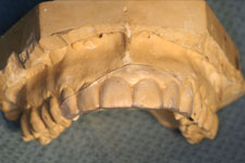
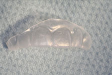
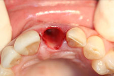
Before surgery, a study model was used to make a "suck down" matrix, used to make the provisional crown. The provisional crown was kept out of occlusion to allow the implant to fully integrate into the patient's bone. In addition, the extraction was performed atraumatically, so that the facial and lingual cortical plates were left intact, and the gingival profile and papillae were preserved.
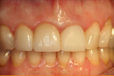
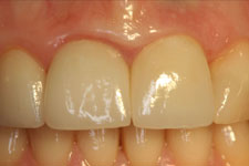
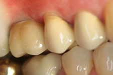
Once the implant was placed, the temporary abutment was seated, and the matrix used to make the screw-retained provisional crown. After implant placement, the patient was seen for post-operative visits at regular intervals to monitor healing and ensure that the gingival contours were being maintained. Once the implant was fully integrated in bone, the patient returned to the restorative dentist for the permanent crown. In this case, the adjacent central incisor also received a new crown, and the end result showed the preserved emergence profile and esthetic crowns.
Pre-Op
|
|
|
|
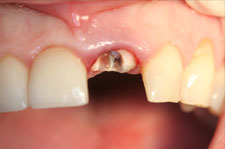
This endodontically treated tooth
has fractured and cannot be
salvaged
|
|
|
|
Important to preserve gingival
contours because of the high smile
line
|
|
|
|
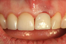
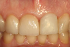
Gingival contours before the
extraction
|
|
|
|
There is more than 3 mm
of bone apical to the root
|
|
At Surgery
|
|
|
|
Study model with matrix
|
|
|
|
Matrix used to make the
immediate provisional crown
|
|
|
|
Flapless and atraumatic extraction
to preserve bone and gingival
tissues
|
|
|
|
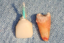
Fractured root with post and core
crown
|
|
At Surgery
|
 |
|
Note alignment and position of first drill in native bone
|
|
At Surgery
|
|
|
|
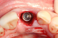
Implant in place in extraction site
|
|
|
|
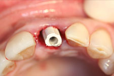
Temporary abutment in place.
|
|
|
|
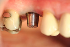
Matrix over temporary abutment, ready to make provisional crown
|
|
|
|
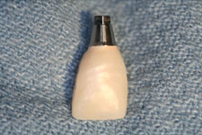
Immediate provisional screw-retained crown, ready to seat.
|
|
|
|
Immediate provisional crown taken out of occlusion
|
|
 |
|
At day of surgery
|
|
Post-Surgery
|
|
|
|
One week after surgery, gingival contours are preserved
|
|
|
|
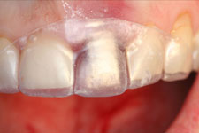
Two months after implant placement, ready to be restored
|
|
|
|
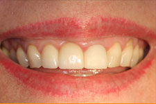
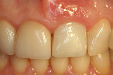
Permanent crown on zirconium
abutment, with new crown on
adjacent central
|
|

|
|
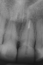
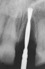
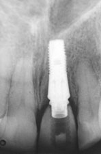
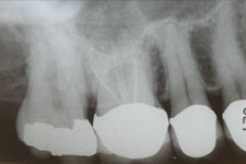
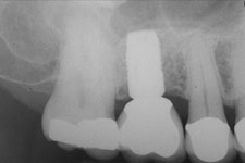
Post-treatment radiograph of final restoration
|
|
Using Trephine Drill and Osteotomes
Placing implants in the posterior areas of the maxilla can sometimes be problematic when there is not enough bone beneath the floor of the sinus to stabilize the implant. Previously, the sequence of treatment started with extraction, then four to six months for the extraction site to heal, followed by sinus augmentation and four to six months additional healing before the implant could be placed - and then more time for implant integration before the patient would finally have the permanent restoration. Using the trephine drill and osteotomes have cut this time considerably.
In the case below, an endodontically treated 1st molar had a fractured root, and the tooth could not be salvaged. An atraumatic extraction preserved the facial and lingual cortical plates; but after four months post-extraction healing, there was not enough bone below the floor of the sinus to place and stabilize the implant.
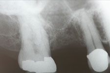
At implant placement surgery, the trephine drill was used to develop a core of bone. This "plug" was then elevated, pushing up the membrane in the floor of the sinus, without penetrating it. The implant was then placed into the prepared site. Because the implant had excellent primary stability, a healing abutment could be placed while the implant integrated. After three months, the implant was ready for restoration. After the final restoration was placed, you will note that new bone has formed completely around the apex of the implant below the sinus membrane.
Pre-Op
|
|
|
|
The root of # 3 is fractured and the tooth cannot be salvaged
|
|
|
|
Four months after the extraction, there is not sufficient bone below the sinus
|
|
|
|
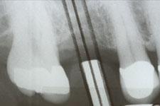
The surgical guide shows alignment for the proposed implant
|
|
|
At Surgery
|
|
|
|
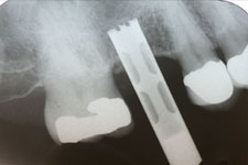
The trephine drill in place at the floor of the sinus
|
|
|
|
The core of bone elevated the sinus membrane, and the implant is in place
|
|
|
|
Implant with healing abutment
|
|
|
Post-Surgery
|
|
|
|
After three months, implant and abutment are ready for restoration
|
|
|
|
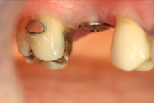
Permanent crown in place.
|
|
|
|
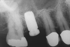
Radiograph of permanent restoration showing bone formed around apex of implant
|
|
|