850 E. Grand Avenue, Suite A
Escondido, CA 92025
Day or night for
24/7 access
7:30 AM to 4:30 PM
Monday through Friday

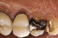
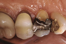
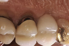
Crown Lengthening
This is a surgical procedure that involves both bone and gingival tissue. From decay or injury, sometimes the crown of a tooth or portions of the crown will break off, leaving insufficient tooth structure for your restoring dentist to make a crown for it. When there is not enough distance between the underlying bone and where the crown will need to be placed, the bone must be contoured to expose more tooth structure. The body needs at least 2 millimeters of distance between where the bottom of the crown is and where the bone is; and while 2 millimeters is a very tiny space, if this distance is not maintained, the body can try to resolve the problem by actually destroying the bone to make enough distance. We will examine the area and decide exactly how much bone will need to be removed, then contour the overlying gum tissue so that your dentist will have a healthy environment and enough tooth structure to make a crown that will last.
|
|
||||||||
|
Frenectomy
Sometimes, there can be a small separation between the two central incisors, leading to a "gap tooth" look. Everyone has a "frenum" - the thick band of tissue that goes between your front teeth; but the place where it attaches and thickness of the tissue varies. When the tissue is thick and the attachment is lower on the gingiva, it can contribute to the separation of the central front teeth, prevent that space from closing during orthodontic treatment, and perhaps even make the space open back up after orthodontic treatment has been completed.
A frenectomy repositions the connection of that band of tissue so that the teeth can come closer together - then stay together permanently. Frenectomies can also be performed for esthetic reasons where orthodontic treatment is not being done. This can reposition the connection of the frenum for those people who might show more of their upper teeth and gums when they smile.
Sometimes the frenum connects on the "inside" or underneath the tongue on the lower arch. This can affect speech, sometimes this is called "being tongue-tied." A simple procedure to reconnect the frenum in a more normal position allows the tongue to have much more freedom to make the sounds associated with normal speech.
Frenectomy |
|||||
|
|
||||
|
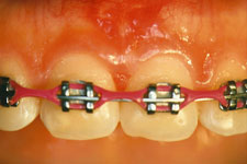
Uncovering Impacted Teeth During Orthodontic Treatment
Any tooth can be "impacted" during growth and development. It just means that the tooth is "hidden" behind bone and/or gingival tissue so that it doesn't erupt or come down into position in the jaw in a normal manner. The most commonly impacted teeth are canines and incisor teeth.
|
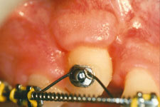
One of the major concerns in uncovering the tooth and allowing it to erupt is to make sure that the tooth erupts into position with the right amount and type of gum tissue around it. There are also different ways of uncovering the tooth, depending on the position of the impaction. Since periodontists are specialists in handling the gingival tissues, we have a few more "tricks" in our tool bag to make sure the final result will include a firm band of pink gum tissue that matches the tissue around the other teeth.
Evaluation and diagnosis of oral diseases
Your dentist may refer you to us so that we can evaluate, diagnose and treat conditions in your mouth. Lichen planus can be very uncomfortable in the mouth, as can Candidiasis (Thrush), a type of yeast infection. There are oral cancers and other benign oral conditions that can make your mouth uncomfortable and also damage your physical health. Please know that when you are referred here for evaluation of your oral condition, we will make sure you are given all information on what we find, what we recommend, and what your treatment options are.
Supportive Care During Radiation and Chemotherapy
Before undergoing head and neck radiation therapy, your radiation therapist may refer you to us for evaluation and recommendations as to what dental treatment needs to be done now, and what supportive care you will need during your radiation therapy. Often, the salivary glands can be temporarily or permanently affected, causing a decreased salivary flow in your mouth. As a consequence, you may have difficulty swallowing, experience a chronic "dry mouth" and also be at risk for cavities. If the density of the jaw bone is expected to be affected, your radiation therapist may want you to have all your dental treatment performed before your radiation therapy begins. We have a great deal of experience with patients in radiation therapy, and we have developed ways to help you control an increase in cavities, ways to help you be more comfortable during and after your radiation therapy, and we can also recommend over-the-counter products that will help. You can also be sure that we will work closely with your radiation therapist to help you through your treatment and keep you as comfortable as possible.
What is "Root Resorption?"
Root resorption is a process that results in loss of tooth structure, somewhat similar to what happens when a tooth decays. The big difference is that this is not caused by bacteria, but by specialized cells in the body (osteoclasts) that mistake the tooth root for bone, and begin the process of dissolving tooth structure instead of destroying and recreating bone, their normal function. We don't yet know why this identification "mistake" occurs; but it is possible to surgically treat the area of resorption by preparing it as if treating an area of tooth decay, and then placing a restoration on the tooth root. Often, this treatment is enough to stop the resorptive process, and the tooth can go on for many years. It is important to understand that sometimes the resorption recurs - again, for some reason the osteoclasts are again fooled into attacking the tooth instead of bone.
When root resorption occurs, it happens under the gingiva, on the part of the tooth root that is in bone. It can be diagnosed through changes in the appearance of the tooth root on radiographs. (If the destructive process has gone into the dentin of the tooth root, then the tooth might also need root canal treatment to return it to health and comfort.) Because this process is taking place under the bone, your periodontist will need to contour the bone so that he can get access to take out the affected area of the tooth root and place the filling. Because he will have to remove bone, he will talk to you about the "crown/root ratio." His recomendation on whether to treat the root resorption will depend on the extent of the resorption, just how much tooth root will be left in bone, and how much of the tooth surface (the crown) will be above the bone - the ratio of the "crown" to the tooth "root." A tooth that can be restored, but will be left with an unfavorable crown/root ratio has a much higher chance of splitting or cracking, as there is not enough root left in bone to withstand biting and chewing pressures.